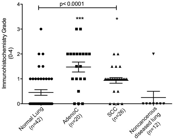
Figure 3.
Immunohistochemical grades of normal lung and NSCLC tissue specimens stained with rabbit polyclonal anti-TMPRSS4. Immunohistochemical staining intensity for each specimen was scored with grades 0–4 (see Materials and methods). Each symbol represents a tumor from an individual patient while the horizontal bars are group mean scores. Statistical comparisons between groups were done using Kruskal-Wallis test (p<0.0001) followed by Dunn’s multiple comparison tests, normal vs adenocarcinoma (***) and normal vs squamous cell carcinoma (*), no significance for normal vs noncancer diseased lung. *p<0.05; ***p<0.001.

