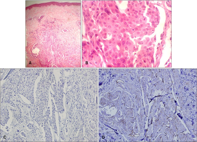
Fig. 2.
(A) Asymmetrical, lobulated cords and nests invading the dermis (H&E, ×40). (B) The large epithelial cells of polyhedral shape showed granular, eosinophilic cytoplasm, large, vesicular, central nuclei and prominent nucleoli (H&E, ×400). (C) The specimen was negative for α-fetoprotein (AFP) (AFP, ×200). (D) CD31 stain did not show any evidence of hematogenous spread (CD31, ×200).

