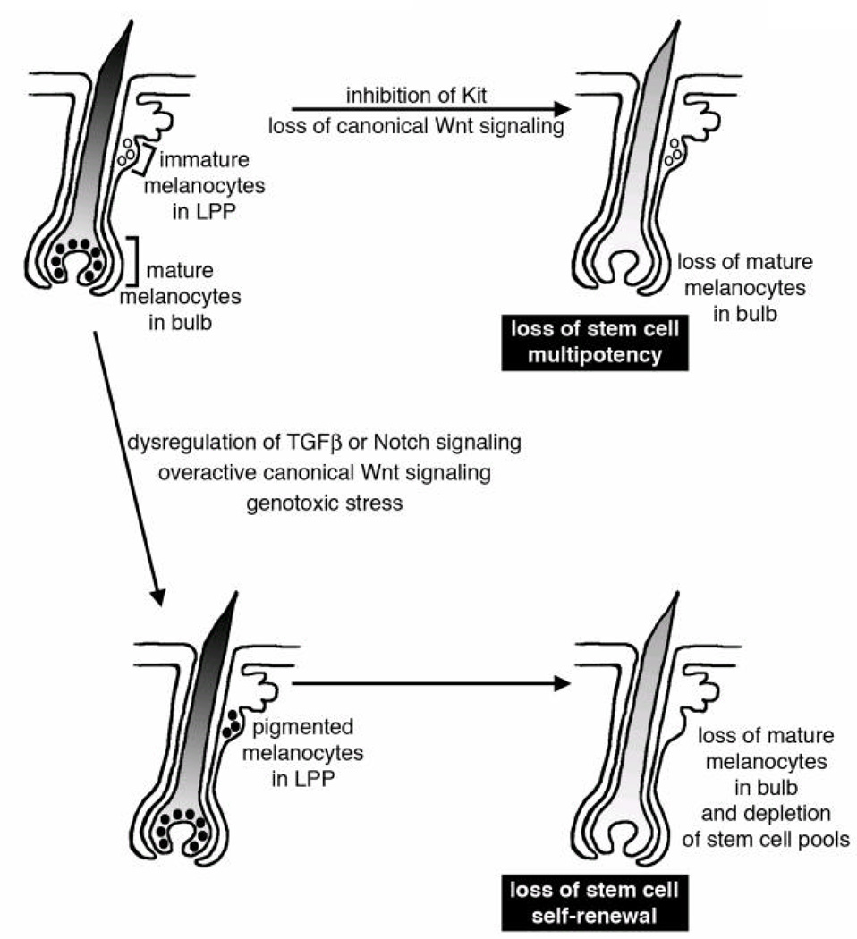
Figure 2. Mechanisms for pigmentation loss and hair graying through extrinsic signals.
In a hair follicle (shown as a schematic for an anagen follicle) immature melanocytes are located in a stem cell niche within the bulge region of the hair follicle, or in a region defined as the lower permanent portion (LPP) (51). Loss of stem cell multipotency occurs when the stem cells fail to differentiate, either permanently or transiently. While immature melanocytes are still found in the LPP, there is a loss of pigmented melanocytes in the bulbar region of the hair follicle. Loss of stem cell multipotency may be caused by inhibiting Kit receptor signaling (52, 53) or through loss of canonical Wnt signaling (56). Loss of stem cell self-renewal occurs when the stem cells prematurely differentiate in the LPP and the stem cell pool is depleted. An error in self-renewal first leads to pigmented melanocytes in the LPP, followed by exhaustion of the stem cells and subsequently by a loss of differentiated melanocytes. A loss of melanocyte stem cell self-renewal may be induced by overactive canonical Wnt signaling (56), dysregulation of TGFβ or Notch signaling (63, 69–72), or through genotoxic stress (124).

