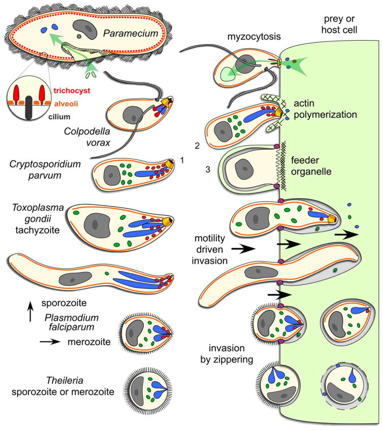
Fig 1.
Schematic comparison of organelles with a role in apicomplexan host cell invasion across the Alveolates. The surface of the free-living ciliate, Paramecium, is covered by cilia which it uses for swimming. Prey bacteria (light green) are taken up through the oral cavity by phagocytosis (green arrow) and merge with lysomes in the cytoplasm for digestion. The enlargement shows the alternating trichocysts (or dense core secretory vesicles (DCSVs)) and cilia underlying the plasma membrane and protruding from the alveolar vesicles. Colpodella vorax is a representative of a dinoflagellate lineage with two flagella that feeds by myzocytosis, also known as cellular vampirism. Note the open conoid structure that is in close apposition upon attachment to a prey cell (flagellate protists). Rhoptries and micronemes are secreted in the process. Prey cell cytoplasm is taken up by pinocytosis and accumulates in basally located vacuoles. Cryptosporidium parvum is an apicomplexan parasite closely related to the archigregarine lineage. A gliding motile sporozoite (1) is shown attaching with its apical end to an endothelial cell of a vertebrate host. Host actin polymerization is induced by the parasite and triggers pseudopod formation, which will engulf the parasite (3), as well as inducing an actin patch, keeping the parasite at the edge of the host cell. This results in extracytoplasmic, yet intracellular, residence of the vacuole. Note the single membrane separating the parasite and host cell cytoplasm, known as the feeder organelle, which is reminiscent of myzocytosis. Toxoplasma gondii tachyzoites and P. falciparum sporozoites display gliding motility, which drives invasion of vertebrate cells. A constriction known as the moving junction forms at the interface of the parasite and the host, and excludes plasma membrane proteins from the host entering into the parasitophorous vacuole membrane. Plasmodium falciparum merozoites enter red blood cells by a combination of gliding motility and zippering. Theileria sporozoites or merozoites are non-motile and can enter host cells in any orientation, a process whereby the dense coat of the zoite is shed. Evidence for micronemes has not been clearly established while no moving junction is formed and the rhoptries are only released upon completion of invasion. Secreted rhoptry and dense granule proteins dissolve the vacuolar membrane and results in cytoplasmic residence. Orange: alveoli or inner membrane complex (IMC); red: micronemes (trichocysts or DSCVs in Paramecium); blue: rhoptries (lysosomes in Paramecium); dark green: dense granules; yellow: conoid. Parasite cells are not drawn to scale.

