Figure 2.

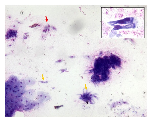
Cytological smears showed, in a mucoid background, scattered histiocytes, gastric and oesophageal mucosal cells, and few groups of ciliated columnar cells (MGG, 200x; inset 400x).

Cytological smears showed, in a mucoid background, scattered histiocytes, gastric and oesophageal mucosal cells, and few groups of ciliated columnar cells (MGG, 200x; inset 400x).