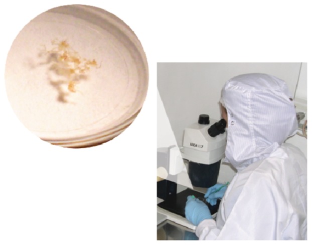
Figure 9.
Photograph showing a petri dish (left) containing seminiferous tubules obtained by microdissection testicular sperm extraction (micro-TESE) immersed in sperm culture medium. The specimen is mechanically minced under stereomicroscopy to release the content of the seminiferous tubules (right).

