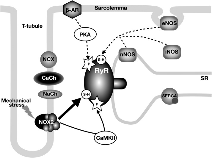
Figure 5.
Signalling pathways involved in regulation of RyR activity in dystrophic cardiomyocytes. The diagram shows the main molecules involved in Ca2+ signalling and EC-coupling. Solid lines depict primary (oxidation and CaMKII-phosphorylation) and dashed lines depict secondary (nitrosation and PKA phosphorylation) mechanisms resulting in hypersensitivity of RyRs. CaCh and NaCh refer to L-type Ca2+ channel and voltage-gated Na+ channel, respectively.

