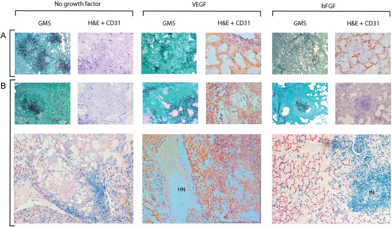
Figure 3.
Angiogenic and inflammatory responses in mouse lungs. Mouse lungs were removed 3 days after infection with Aspergillus fumigatus and stained with Gomori methenamine silver (GMS) or hematoxylin and eosin (H&E) combined with CD31 immunostaining, as described in Methods. Lungs of saline-treated mice (A) and amphotericin B (AmB)–treated mice (B) showed typical features of invasive aspergillosis in neutropenic hosts, consisting of extensive mycelial invasion of lung parenchyma and scant neutrophilic infiltrate. CD31-positive microvessels were absent from the periphery of fungal lesions. In both recombinant human vascular endothelial growth factor, proximal splice site 165 (VEGF)–treated mice and recombinant human basic fibroblast growth factor (bFGF)–treated mice, fungal hyphae were more widely dispersed, and networks of CD31-positive microvessels were observed around and inside Aspergillus-infected zones. Angiogenesis was disordered in AmB/VEGF-treated lungs, and large areas of hemorrhagic necrosis (HN) were observed. In AmB/bFGF-treated mice, the angiogenic response seen in bFGF-treated mice was augmented by neutrophilic infiltration of Aspergillus-infected zones, inflammatory necrosis (IN), and reduced fungal burden. Magnification: GMS, ×100; H&E/CD31, ×400; bottom panel, ×100.

