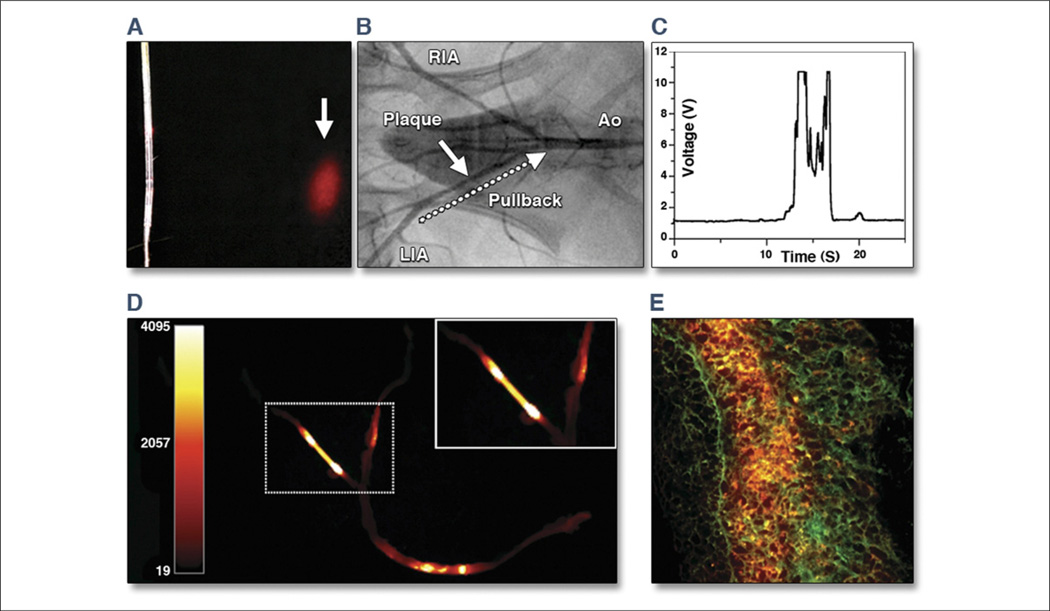
Figure 11. Real-Time Intravascular NIRF Imaging of Protease Activity.
(A) The intravascular catheter was modified from a clinical optical coherence tomography guide wire used in human coronary artery imaging. Near-infrared fluorescence (NIRF) (red) was emitted in a 90° arc and focused 2 mm away from the aperture. (B) Angiogram of atherosclerotic iliac arteries after balloon injury and hyperlipidemic diet. White arrow indicates plaque. Twenty-four hours after Prosense750 (VisEn Medical, Woburn, Massachusetts) injection, the NIRF guide wire was placed percutaneously into the left iliac artery (LIA) and then pulled back (dotted arrow) manually over 20 s. Ao = aorta; RIA = right iliac artery. (C) Real-time voltage recordings of NIRF showed signal peaks in areas of plaques but not in control segments. (D) Ex vivo fluorescence reflectance images corroborated the in vivo imaging findings. (E) Merged 2-color fluorescence microscopy of atheroma section demonstrated focal plaque NIRF (orange) that was spatially distinct from 500 nm autofluorescence (green). Modified with permission from Jaffer et al. (147).

