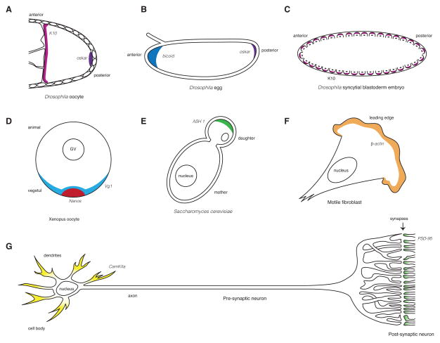
Figure 1. mRNA localization in Drosophila oocytes and embryos, and animal neurons.
A. K10 mRNA (magenta) localizes to the anterior and oskar mRNA to the posterior (purple) of a developing Drosophila oocyte. The oocyte is depicted on the right, surrounded by somatic follicle cells. B. bicoid mRNA (blue) localizes to the anterior and oskar (purple) mRNA to the posterior of the Drosophila egg. C. K10 mRNA (magenta) is found at the apical side of the blastoderm nuclei (black), which are partially separated from one another by invaginated cell membranes. D. Nanos (red) and Vg1 (pale blue) localize to the vegtal pole of the Xenopus oocyte utilizing two alternative localization pathways referred to as the early (active in stages I and II of oogenesis) and late (active in stages II–IV) pathways, respectively. The oocyte depicted is in stage IV of oogenesis. E. ASH1 mRNA (green) localizes to the distal tip of a budding daughter cell during mitosis in the yeast Saccharomyces cerevisiae. F. β-actin mRNA (orange) localizes to the leading edge of motile fibroblast. G. CamKIIα mRNA (yellow) localizes to the dendrites of a neuron, whereas PSD-95 mRNA (pale green) is found at the post-synaptic side of a synapse.

