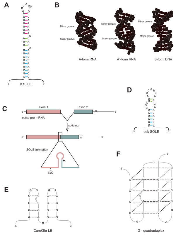
Figure 2. Structural motifs involved in mRNA localization.
A. Secondary structure of the K-10 LE. The three helical sections of the hairpin are shown with colored base-pairs (proximal blue, medial green, and distal pink). A flipped out U is present in the loop. This structural representation is based upon Figure 1 of [30••]. B. Depiction of A-form RNA, A′-form RNA, and B-form DNA helices. The K-10 LE stem folds into A′-form helices, in which the size of the major groove is similar to that of B-form DNA. This is in contrast to the small major groove found in the more common A-form RNA helix. This image is again based upon Figure 1 of [30••]. C. The SOLE is formed after splicing of the first intron in osk pre-mRNA [32••]. Exon 1 is shown as pink and exon 2 is green in both the pre-mRNA (top) and the spliced product (below). The inset shows the exon 1 (pink) and exon 2 (green) sequences within the SOLE and the position of the EJC (open arrowhead), 20–24nts upstream of the splice junction (filled arrowhead). D. The oskar SOLE is predicted to form a hairpin, with the proximal and distal stems shown with blue and green basepairs, respectively. E. Secondary structure of the CamKIIα LE in mammals, as depicted in [37•]. F. CamKIIα LE folds into a G-quadruplex through hydrogen-bond interactions indicated by dashed lines.

