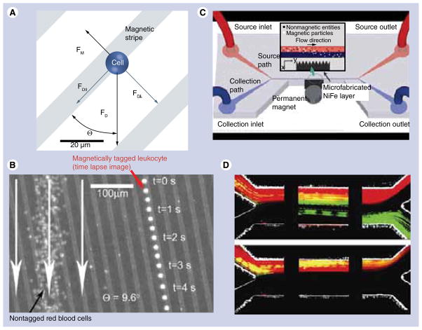
Figure 9. Cell sorting in magnetic microfluidic chips.
(A) Principle of cell separation by combining the hydrodynamic force with magnetic field gradient provided by ferromagnetic nickel stripes. The cells are coupled with superparamagnetic iron oxide beads to enable magnetically assisted sorting. (B) Overlay of optical reflection image of the microfluidic magnetic chip and the fluorescent images of a single magnetically labeled leukocyte (right) and a large number of free-flowing red blood cells (left). The image demonstrates that the magnetically tagged cell is trapped between magnetic stripes and it follows the defined path in contrast with untagged cells. (C) Schematic diagram of the cell sorting device with microfluidic channel and microfabricated permalloy comb separated spatially for improved biocompatibility. (D) Separation of green fluorescent magnetic beads and red blood cells labeled with a red fluorescent dye SYTO 64. The snapshots represent the distribution of cells/beads mixture as it enters the channel (left), proceeds to the middle (middle) and separates at the end of the channel (right). The images were acquired with (top) and without (bottom) the permanent magnet, which is used for magnetizing the permalloy comb. In the absence of the external magnetizing field no separation is observed (bottom).
(A & B) Reproduced with permission from [72].
(C & D) Reproduced with permission from [76].

