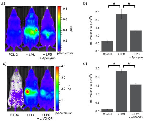
Figure 9. Bioluminescent signal from PCL-2 and IETDC following lipopolysaccharide challenge.
(a) Representative image (10 min post-injection) of mice 6 h after injection of LPS (IP, 3 mg/kg in 50 μL of saline) or saline vehicle (IP, 50 μL). Two minutes prior to imaging, mice were treated with apocynin (IP, 10 mg/kg in 20 μL of DMSO) or vehicle (IP, 20 μL DMSO). Imaging was started following injection of a mixture of PCL-2 and D-cysteine (IP, 0.05 μmol each, in 50 μL of 1:1 DMSO:PBS). (b) Total photon flux, 0–15 min post-injection, for mice injected with PCL-2 and D-cysteine, ± LPS, and ± apocynin. (c) Representative image (30 min post-injection) of mice 6 h after injection of LPS (IP, 3 mg/kg in 50 μL of saline) or vehicle (IP, 50 μL saline). Thirty minutes prior to imaging, mice were treated with z-VD(OMe)-OPh (IP, 1 μmol in 20 μL of DMSO) or vehicle (IP, 20 μL DMSO). Imaging was started following injection of a mixture of IETDC and HCBT (IP, 0.05 μmol each, in 50 μL of 1:1 DMSO:PBS). (d) Total photon flux, 15–45 min post-injection, for mice injected with IETDC and HCBT, ± LPS, and ± z-VD(OMe)-OPh. Statistical analyses were performed with a two-tailed Student’s t-test. *P < 0.05 (B and D: n = 3–4) and error bars are ±SEM.

