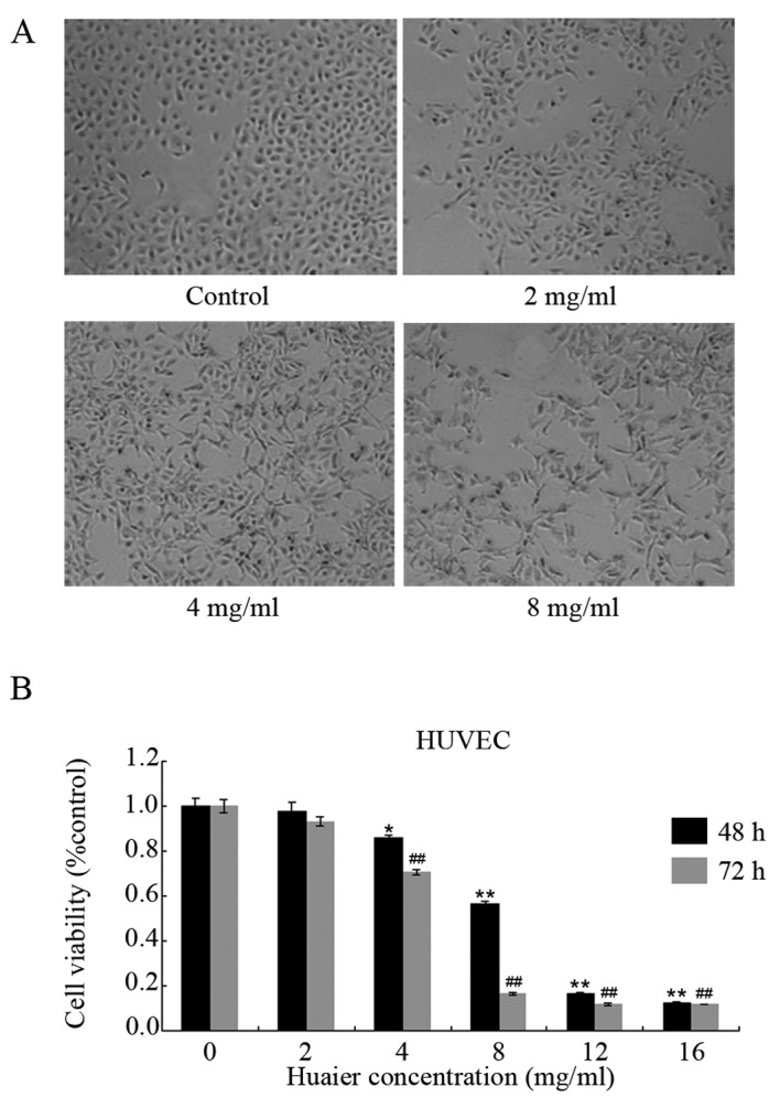
Figure 1.

Morphological and viability changes in HUVECs induced by Huaier extract. (A) HUVECs were seeded in 24-well plates and incubated with Huaier extract for 24 h. Representative images of HUVECs in the control and treated groups. (B) Huaier extract inhibited cell proliferation in a time- and dose-dependent manner. The results are presented as the means ± SD of 3 independent experiments conducted in triplicate. *P<0.05; **P<0.01; #P<0.05; ##P<0.01.
