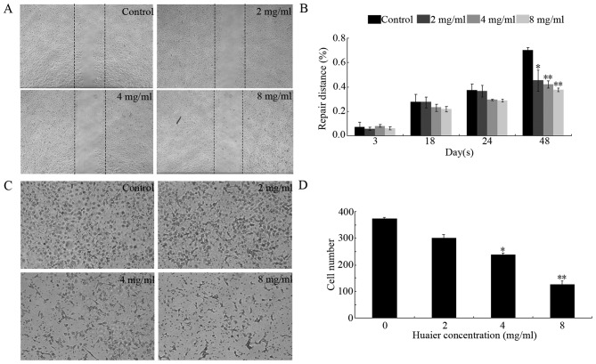
Figure 3.
Effect of Huaier extract on the cell migration of HUVECs. (A) Confluent monolayers of HUVECs on a 12-well plate were wounded using a pipette tip and treated with Huaier extract or the vehicle. The images of wound closure were captured under a phase-contrast microscope after 48 h. (B) The migration inhibition is presented as distances between the 2 edges of the scratch. (C) Chemotaxis assay of HUVECs, which shows the inhibitory effect of Huaier extract on cell migration following 24 h of treatment. The cells successfully migrated to the lower surface of the insert. (D) The cell numbers decreased in a dose-dependent manner following treatment with Huaier extract. In the histograms, all values are expressed as the means ± SD. *P<0.05; **P<0.01 as compared with the vehicle.

