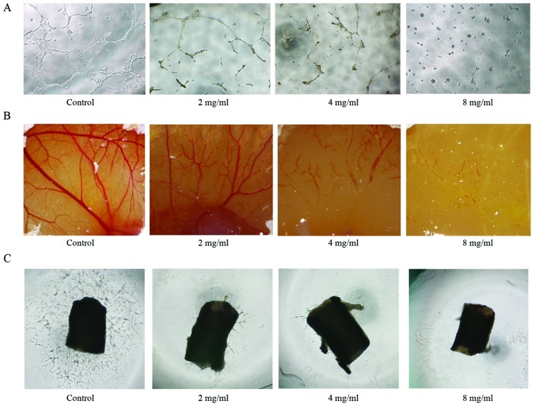
Figure 4.
Huaier inhibited angiogenesis in vitro and ex vivo. (A) HUVECs were seeded on Matrigel-coated 96-well plates and incubated in the absence or presence of Huaier extract for 9 h. Representative images of HUVEC tube formation. (B) CAM of 9-day-old chick embryos exposed to Huaier extract or the vehicle. After 24 h of incubation, the CAM tissue directly beneath each filter disc was photographed. The image represents at least 6 chick embryos. (C) Examples of aortic rings of mice fed with DMEM containing 20% FBS in the control and treated groups. Representative images of vessel sprouting were taken at day 5 of treatment.

