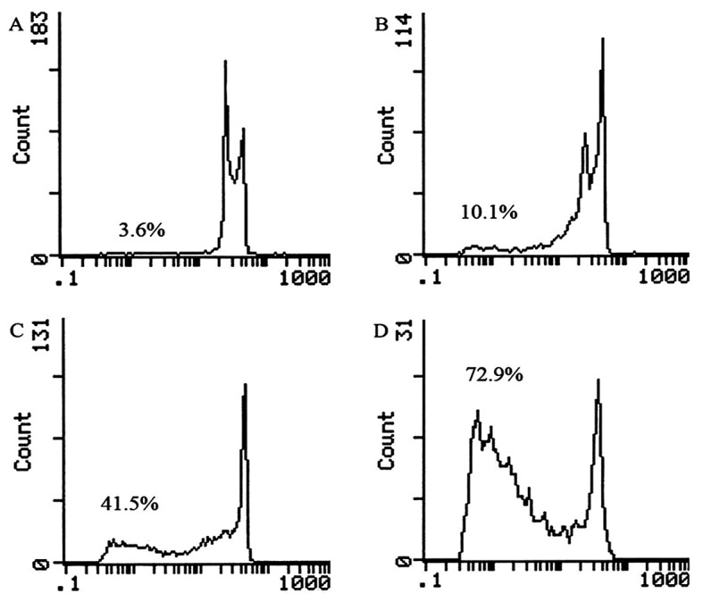
Figure 4.

Representative DNA fluorescence histograms of propidium iodide (PI)-stained cells. MPC-11 cells were (A) untreated or treated with various doses of CPT-TMC [(B) 12.5 ng/ml, (C) 25 ng/ml, (D) 50 ng/ml] for 48 h. The cells in the sub-G1 phase were considered as apoptotic cells. The apoptosis rates in non-treated and CPT-TMC-treated cells were (A) 3.6%, (B) 10.1%, (C) 41.5%, (D) 72.9% as assessed by flow cytometry.
