Figure 5.

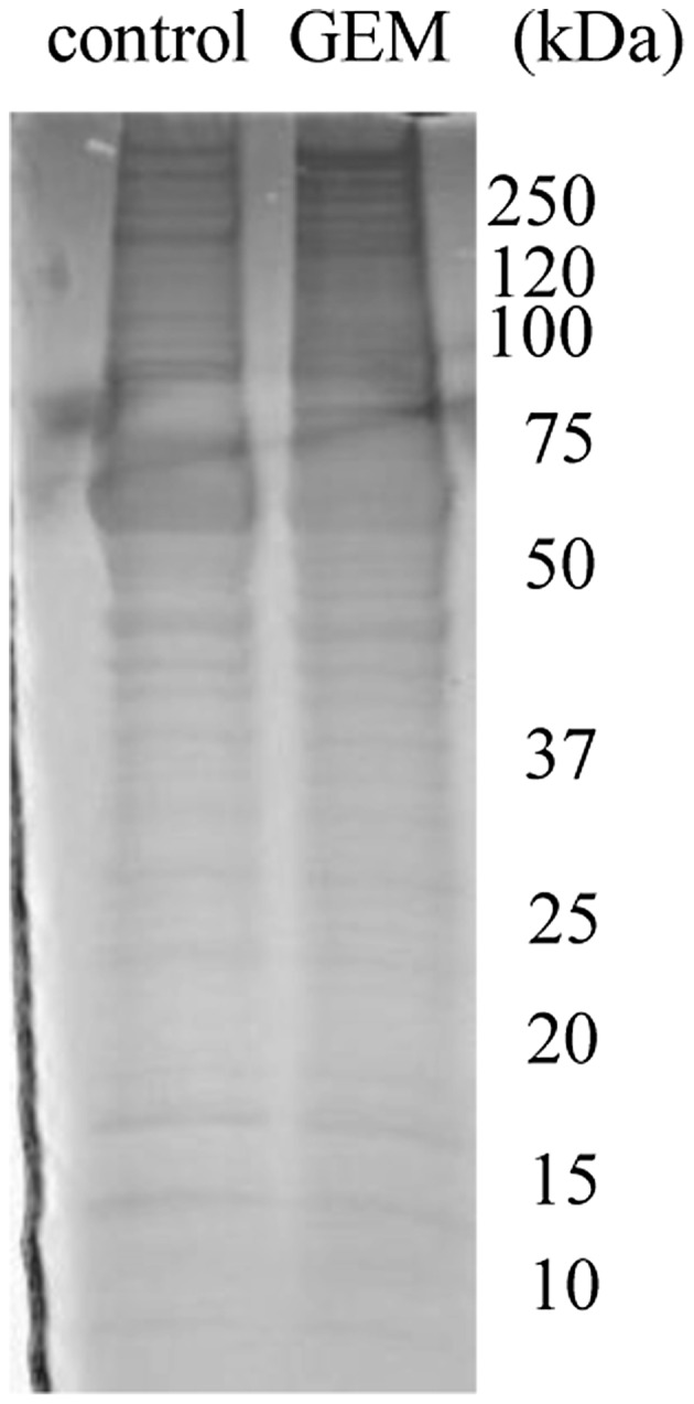
SDS-PAGE analysis of CM. Fifty micrograms of protein was applied to the gel, and the gel was stained with Coomassie Brilliant Blue (n=3). In the regions of 60–8 and 20–30 kDa, control and treatment samples were of equal quantity.

SDS-PAGE analysis of CM. Fifty micrograms of protein was applied to the gel, and the gel was stained with Coomassie Brilliant Blue (n=3). In the regions of 60–8 and 20–30 kDa, control and treatment samples were of equal quantity.