Fig. 1.
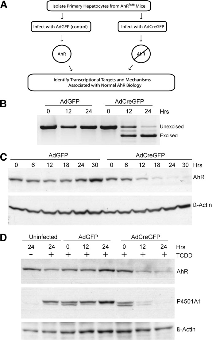
Monitoring loss of the AhR in primary hepatocytes. (A) Schematic depicting the general strategy used to generate AhR-positive and AhR-negative primary hepatocytes. (B) RT-PCR was performed to monitor excision of the AhR gene loxP-flanked exon 2 with use of total RNA isolated from AhRfx/fx primary hepatocytes infected with AdGFP (control) or AdCreGFP (Cre recombinase) for the indicated times. PCR primers were designed to amplify different size PCR products distinguishing the unexcised from excised transcript. (C) Whole-cell lysate from AhRfx/fx primary hepatocytes infected with AdGFP or AdCreGFP were analyzed using Western blotting for AhR protein. Actin was used as a loading control. (D) AhRfx/fx primary hepatocytes were either uninfected or infected with AdGFP or AdCreGFP for the indicated times, followed by treatment with DMSO (lane 1) or 6 nM TCDD (lanes 2–8) for 6 hours. AhR and P4501A1 protein expression was monitored using Western blot analysis. Actin was used as a loading control.

