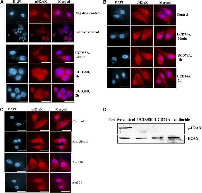
Fig. 12.
H2AX phosphorylation. U87MG glioma cells were treated with drugs UCD38B, UCD74A, or amiloride (250 μM). U87MG cells treated with staurosporine (1 μM) were used as positive control to demonstrate H2AX phosphorylation. Immunostaining (A, B, and C) of γH2AX in treated glioma cells was detected using anti-γH2AX polyclonal antibody. (D) Immunoblot of U87MG cells treated with staurosporine, UCD38B, UCD74A, or amiloride. Anti-γH2AX antibody was used to detect phosphorylated H2AX that was normalized to H2AX and detected using anti-H2AX antibody. Only H2AX was seen in treated cells except for staurosporine (positive control), which demonstrated γH2AX.

