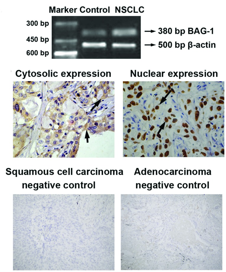
Figure 1.
mRNA and protein expression of BAG-1 in lung tissues of NSCLC patients in I–IIIA stage. (A) Total RNA was isolated from lung tissues and RT-PCR was performed. Agarose gel eletrophoresis of PCR products were shown as indicated. Maker DL1000 was used to indicate PCR product lengths. Lung tissue samples obtained from healthy subjects were applied as control. (B) The 10% formalin-fixed and paraffin-embedded lung tissue sections were stained with anti-BAG-1 antibody. Immunoreactivity was visualized using an SP immunohistochemical staining kit according to the manufacturer's instructions. Immunohistochemical analysis showed cytosolic and nuclear expressions of BAG-1 in lung tissues of NSCLC patients. Negative controls for specimens obtained from patients with squamous cell carcinoma and adenocarcinoma are presented.

