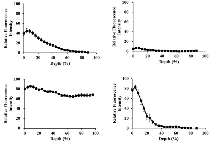
Figure 2.
Distribution of four different compounds in MCLs of DLD-1 cells following 2 h of exposure. MCLs were exposed to (A) 40 μM of calcein-AM, (B) 50 μM of PTX-rd, (C) 100 μM of DOX, and (D) 20 μM of ethidium homodimer-1 (EthD-1). Exposure to calcein-AM occured at room temperature, whereas others took place at 37˚C.

