
Figure 4.
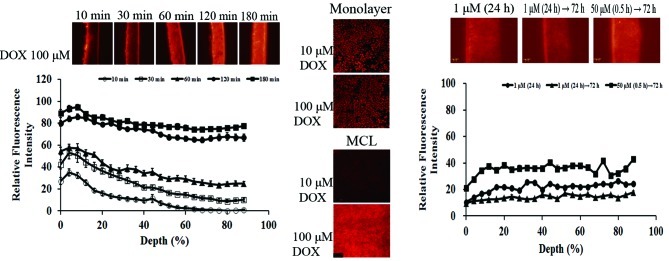
Distribution of DOX in MCL under various exposure conditions. (A) MCL were exposed to 100 μM of DOX up to 3 h. Drug exposure was at the top side of the MCL (0% depth). (B) Monolayers and MCL were exposed to DOX (10 and 100 μM) for 3 and 4 h, respectively. Confocal section of MCL was taken at 90 μm (60% depth) distance from the top. (C) MCL were exposed to 1 μM for 24 h or 50 μM for 30 min, then further incubated in drug-free media until 96 h. Drug exposure was at the bottom side of the MCL (100% depth). In (A) and (C), fluorescence within the bottom 10% was deleted in order to eliminate over-spill from the membrane.