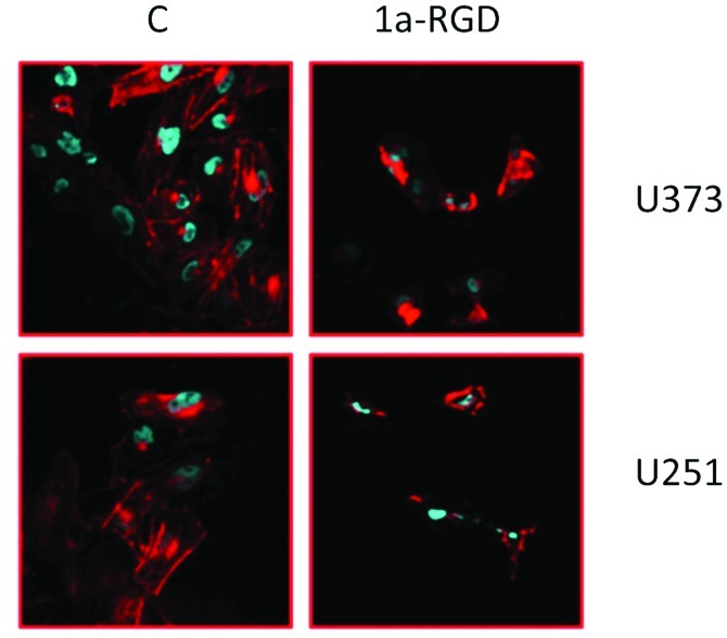
Figure 7.
Cytoskeleton disassembly. Cells were plated onto polylysine coated coverslips and incubated in the presence of 20 μM 1a-RGD for 4 h. At the end of the treatments, the cells were stained with fluorescent phalloidin and Hoechst 33342. Images were acquired with laser scanning confocal microscopy. Each experiment was repeated at least twice.

