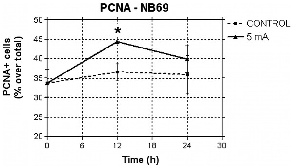
Figure 5.

Immunofluorescence assessment of PCNA expression in NB69. Percents of PCNA-positive cells before (t = 0 h), during (t = 12 h) and the end (t = 24 h) of treatment with 50 μA/mm2. The fraction of cells undergoing DNA synthesis increased significantly during treatment. Approximately 23,000 cells were counted in a total of four experimental replicates. Approximately 5,000 cells per experimental group were evaluated. Means ± SEM; *0.01< p<0.05, Student's t-test.
