
FIGURE 3:
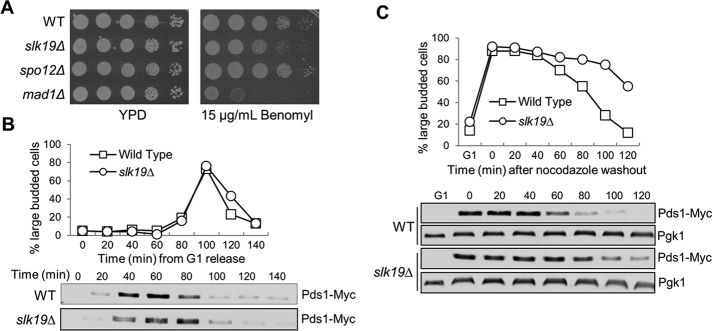
slk19 mutant cells show delayed anaphase entry after nocodazole treatment. (A) slk19Δ mutants show sensitivity to benomyl. WT, slk19Δ, spo12Δ, and mad1Δ mutant cells were grown to saturation, 10-fold diluted, and spotted onto YPD with and without 15 μg/ml benomyl. The plates were scanned after incubation at 30°C for 3 d. (B) slk19Δ mutants do not show an obvious cell cycle defect in an unperturbed cell cycle. WT and slk19Δ cells with PDS1-18myc were released from G1 into YPD at 25°C. Cells were harvested every 20 min and examined for their budding index and Pds1 protein levels. (C) slk19Δ mutants show an anaphase entry delay after exposure to nocodazole. G1-arrested WT and slk19Δ cells with PDS1-18myc were released into YPD containing 20 μg/ml nocodazole at 25°C for 120 min. Nocodazole was then washed off, and the cells were released into YPD medium at 25°C. Cells were harvested every 20 min for budding index and Pds1 protein levels. Pgk1 protein level is shown as a loading control.