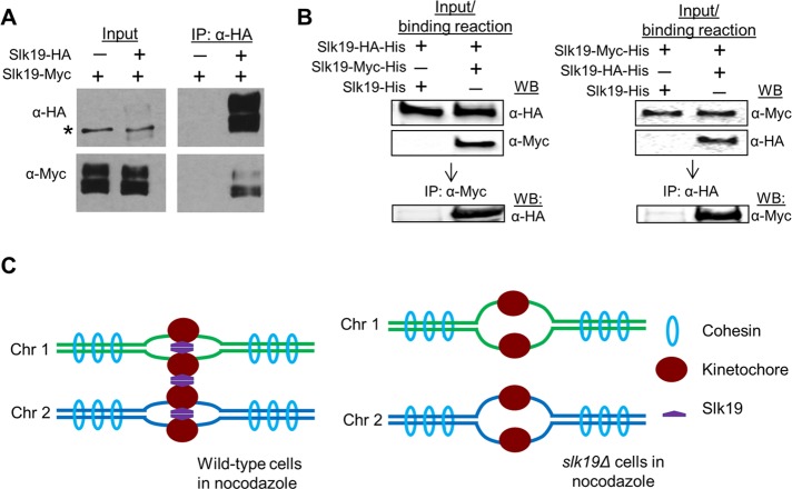
FIGURE 6:
Slk19 protein forms dimers. (A) Both full-length and cleaved fragment of Slk19 physically interact with themselves. Diploid yeast strains containing either SLK19/SLK19-13Myc or SLK19-6HA/SLK19-13Myc were grown to log phase. The cells were harvested to prepare the whole cell extracts with a bead beater. The extracts were first incubated with anti-HA antibody, and then protein A/G-coated agarose beads were added. Western blotting was performed to detect Slk19-6HA and Slk19-13Myc in the whole-cell extracts and the immunoprecipitates. Asterisk indicates a nonspecific band. (B) Slk19 interacts with itself in vitro. Purified Slk19-HA-His was mixed with Slk19-His or Slk19-Myc-His, and then the mixtures were pulled down with anti-Myc antibody. Slk19 proteins with different tags in the mixture and immunoprecipitates were detected using Western blotting with anti-Myc and anti-HA antibodies. A similar pull-down assay was performed after the mixture of Slk19-Myc-His with Slk19-His or Slk19-HA-His. (C) The working model for Slk19-dependent kinetochore clustering and sister kinetochore cohesion.

