
FIGURE 8:
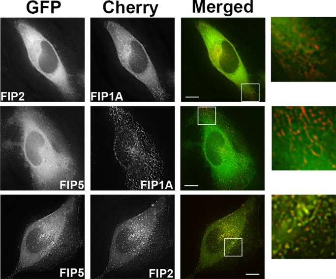
FIP1A resides on distinct compartments from FIP2 and FIP5. Time-lapse imaging of live HeLa cells transfected with EGFP-FIP2 or EGFP-FIP5 (Supplemental Video S10) and mCherry-FIP1A was conducted with deconvolution microscopy. FIP1A was found on compartments separate from FIP2 and FIP5 in peripheral regions of the cells. Time-lapse imaging of live HeLa cells transfected with EGFP-FIP5 and mCherry-FIP2 (Supplemental Video S11) was also conducted, and FIP5 and FIP2 overlapped both in the perinuclear and peripheral regions. Data are representative of at least three independent experiments. Bars, 10 μm.