
Figure 1.
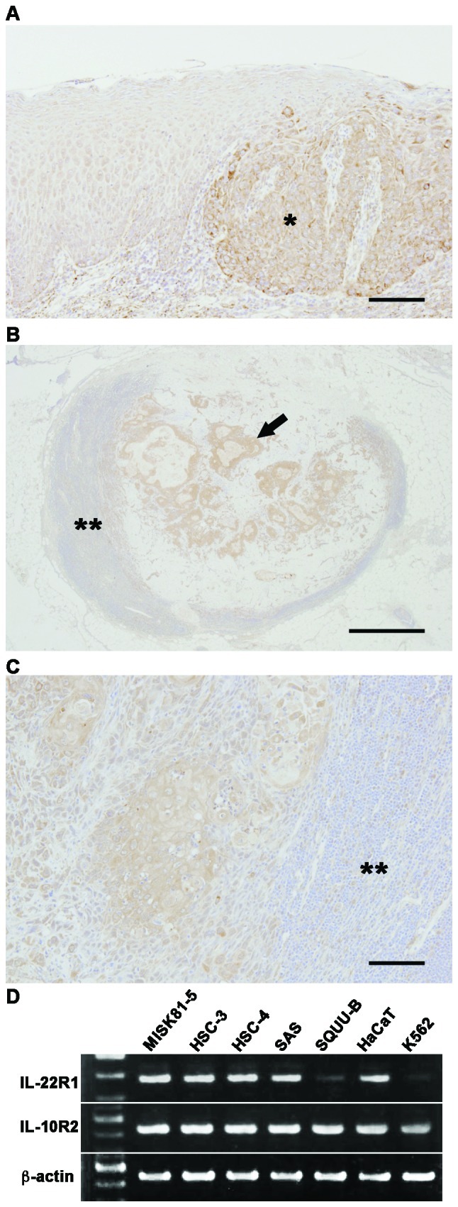
IL-22R is expressed in the OSCC of the extirpated samples and OSCC cell lines. (A) IL-22R immunostaining was observed in four of the seven extirpated samples, including some normal regions. The immunoreactive intensity for IL-22 was increased in the OSCC region (right side; *) compared with that in the normal oral epithelium (left side). Scale bar, 100 μm. (B) Metastatic carcinoma cells in the cervical lymph node (**) showed strongly positive signals for IL-22R (arrow). Scale bar, 1 mm. (C) IL-22R positive signals in metastatic carcinoma cells in the cervical lymph node (**) in another case. Scale bar, 100 μm. (D) IL-10R2 and IL-22R1 mRNA expressions were examined by semiquantitative RT-PCR. β-actin was used as an internal control.