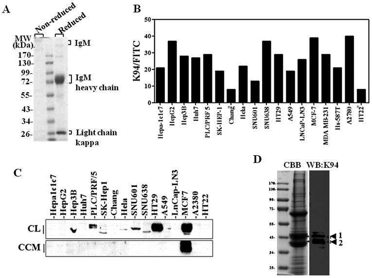
Figure 1.
Target antigen of K94 tumor-associated autoantibody in various human tumor cell lines. (A) Purified K94 monoclonal antibody separated on non-reducing or reducing 10% SDS-PAGE and stained with Coomassie Blue. (B) FACS analysis of intracellularly stained tumor cell lines with K94 antibody. (C) Western blot analysis of target antigen of K94 autoantibody. Total cell lysates (CL, 50 μg) or concentrated cell culture media (CCM, 50 μg) were resolved on 8–10% SDS-PAGE gels, followed by blotting and probing with K94 autoantibody. (D) Fractions enriched with K94 autoantigen (described in the Materials and methods, in detail) were pooled, concentrated and resolved on a 10% SDS-PAGE gel. Two protein bands corresponding to K94 autoantigen confirmed by immunostaining with K94 antibody were excised and analyzed by proteomic methods.

