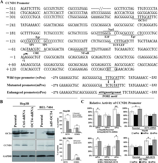
Figure 3.
CCND1 promoter activity in HCC cells. (A) The wild type CCND1 promoter (wPro) containing the transcription factor binding sites was cloned from HepG2 DNA genome. From the wPro, the motif-mutated promoter (mPro) and motif-enhanced promoter (ePro) within −252 to −245 were amplified and used to construct the CCND1 promoter-driven luciferase plasmids, pGL3wPro-Luc, pGL3mPro-Luc and pGL3ePro-Luc. (B) CCND1 and OCT4 expression was detected by Western blotting in HCC cells. GAPDH was used as the loading control, and densitometry analysis was performed to show CCND1 and OCT4 expression levels normalized with GAPDH density; **, P < 0.01. (C) The indicated parental, adenovirus-infected and shRNA-transfected cells were seeded on 24-well plates at a density of 5 × 10⁴ cells/well and transfected with the plasmids pGL3wPro-Luc, pGL3mPro-Luc and pGL3ePro-Luc at 200 ng/well. The relative activity of CCND1 promoter in HCC cells was measured and shown in histograms; *, P < 0.05; **, P < 0.01.

