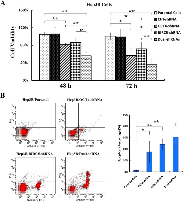
Figure 4.

Cell growth inhibition and cell apoptosis induced by co-suppression of OCT4 and BIRC5 in HCC cells. (A) The parental and indicated shRNA-transfected Hep3B cells were cultured in 96-well plates at a density of 5 × 10³ cells/well for 48 and 72 h. Cell viability was measured by MTT assay at a wavelength of 570 nm with a reference wavelength of 655 nm and shown in histograms; *, P < 0.05; **, P < 0.01. (B) The parental, adenovirus-infected and shRNA-transfected Hep3B cells at 106 cells/ml were stained with PI and Annexin V-FITC for detection of cell apoptosis. Percentages of cell apoptosis were shown in histograms; *, P < 0.05; **, P < 0.01.
