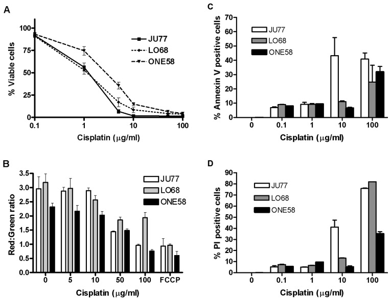
Figure 1.
Mechanisms of cisplatin induced cell death in mesothelioma cells. (A) In vitro sensitivity of MM cell lines to cisplatin. Cells were cultured in the presence of cisplatin for 24 h. Cell viability was determined by MTT assay. Data presented are the mean ± SD of five independent experiments each performed in triplicate. (B) Dose-dependent mitochondrial membrane depolarisation. Mitochondrial membrane potential was measured by JC-1 accumulation following cisplatin treatment (lower ratio of red/green indicates loss of potential). Results are the mean ± SD of at least three independent treatments performed in triplicate. FCCP-positive control. (C) Phosphatidylserine (PS) translocation in response to cisplatin. PS translocation to the outer cell membrane was measured by Annexin-V binding and flow cytometry. Cells were treated with a range of cisplatin concentrations (0.1–100 μg/ml) for 24 h. Results shown are the mean ± SD of two independent experiments performed in triplicate. (D) Loss of membrane integrity in response to cisplatin. Membrane integrity was measured by staining with the cell impermeant dye propidium iodide and flow cytometry. Results shown are the mean ± SD of two independent experiments performed in triplicate.

