
Figure 3.
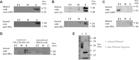
The β3 Ig domain binds β3 and β1. A–C) HEK293 cells were cotransfected with plasmids expressing β3-Ig-6His and β3-wt-EGFP(A); β1-wt (B), or β3-ΔECD-EGFP (C). Lysates were precipitated with Talon (anti-His) beads and blotted with antibodies as indicated. D) Control lanes showing cells transfected with only β3-Ig-6His together with untransfected cells. Lysates were precipitated with Talon (anti-His) beads and blotted with antibodies as indicated. FT, flow through; W, wash; E, elution fraction. E) PNGase F digestion reduces the higher-molecular-mass doublet characteristic of β3-Ig-6His protein to a single band of 15–16 kDa. Mwt, molecular weight (mass). Asterisk indicates the added PNGase F enzyme.