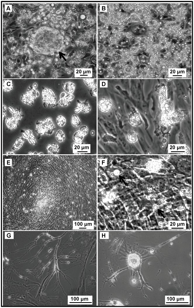
Figure 2. Ascites-derived chicken ovarian cancer (COVCAR) cells in culture.
COVCAR cells were harvested from ascites and cultured as described in Materials and Methods section. Notice several translucent vesicles in the cytoplasm and spherical shape of the cells (A, B, C, and D). A few large spheroid mass of COVCAR cells were noticed as shown in A (arrow). Many cells had papilla-like or microvilli-like projections on the cell surface (C). Notice fibroelastic transformation of spherical COVCAR cells (D). E-F. Multiple layers of fully confluent COVCAR cells appearing as an interwoven mat. A few floating cells as shown in F (arrows) displayed translucent vesicles in the cytoplasm. G. COVCAR cells appearing as a network of tube-like structure. H. A few senescent cells showing cellular hypertrophy and stellate appearance.

