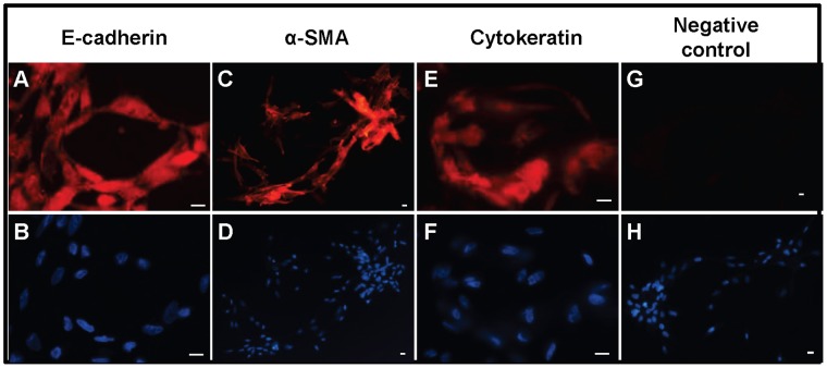
Figure 6. Expression of cytoskeletal proteins in chicken ovarian cancer (COVCAR) cells.
Photomicrographs of chicken ovarian cancer (COVCAR) cells showing E-cadherin (A), α-smooth muscle actin (C; SMA), and cytokeratin (E) immunostaining. Paraformaldehyde-fixed COVCAR cells were immunostained as described in Materials and Methods section. Fixed COVCAR cells were incubated with anti-mouse IgG (G) in place of primary antibody as negative control. Nuclei were visualized with DAPI staining (B, D, F, and H) on cells immunostained with E-cadherin, SMA, or cytokeratin, respectively. Scale bars-10 µm.

