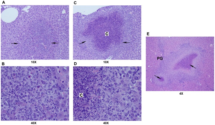
Figure 3. Pyogranulomatous inflammation of liver.
Sections with varying degrees of severity of pyogranulomatous lesions: in (A) and (B), focal pyogranuloma (arrows) without caseous necrosis (shown at 10 and 40X magnification, respectively); in (C) and (D), a larger pyogranuloma (arrows) with a caseous necrotic center (designated as C; shown at 10 and 40X magnification, respectively); and in E, confluent pyogranulomas with caseous necrotic centers (arrows; shown at 4X magnification). Details of processing and staining of the tissue are described in Materials and Methods.

