
Figure 4. Pyogranulomatous inflammation of the spleen.
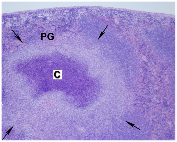
Cross section showing pyogranulomatous inflammation (PG,) wherein arrows delineate borders of the pyogranuloma. The center has caseous necrosis (designated by C; shown at 4X magnification). Details of processing and staining of the tissue are described in Materials and Methods.