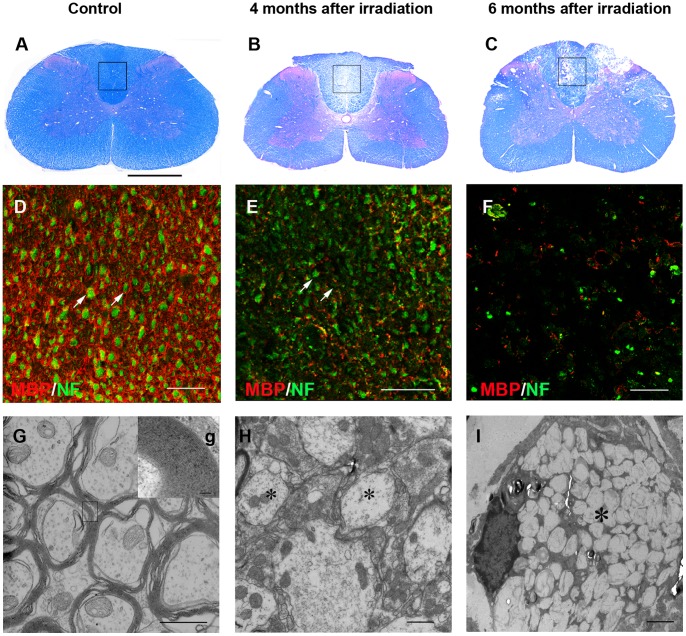
Figure 1. Radiation-induced demyelination in the dorsal funiculus of cervical spinal cord.
(A) LFB/H&E staining showed normal spinal cord without irradiation. (B) Four months after irradiation, there was a focal demyelinated zone in the dorsal funiculus. (C) Six months following injury, focal necrosis was seen in the dorsal funiculus. (D) Immunostaining for MBP and NF, enlargement of framed area in A, depicted that non-irradiated myelin surrounding the axons (arrows). (E) Four months after irradiation, most of the axons lost myelin (arrows). (F) At six months, axons began to show necrosis. (G) Electron microscopy confirmed the normal structure of myelin. g, Higher magnification of the tissue in the box in G, indicated the compact myelin sheath. (H) Four months after irradiation, most of axons remained demyelinated (asterisk). (I) Six months following damage, axons necrosis was found (asterisk). Bar, 200 µm (A–C); bar, 50 µm (D–F); bar, 1 µm (G, H); bar, 100 nm (g); bar, 2 µm (I).

