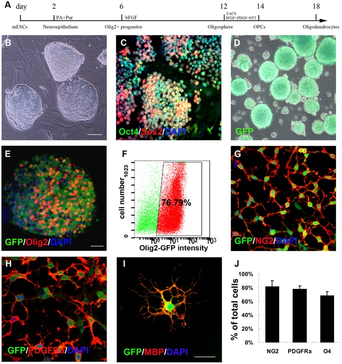
Figure 2. In vitro differentiation of Olig2-GFP-mouse mESCs into Olig2+-GFP+- OPCs.
(A) Schematic procedure for differentiation of Olig2-GFP-mESCs into Olig2+-GFP+-OPCs. (B) Phase contrast photograph of mESCs colonies on mouse embryonic fibroblast. (C) mESCs remained undifferentiated and stained positive for Oct4/Sox2. (D) Phase contrast photograph of Olig2+-GFP+ spheres at day 12. (E) GFP+ spheres expressed Olig2. (F) At day 12, the percentage of GFP+ cells was 76.79%±1.35%, sorted by FACS method. At day 14, all purified OPCs expressed GFP as well as the OPC markers NG2 (G) and PDGFRα (H). At day 18, plated cells adopted a typical oligodendrocyte morphology characterized by multiple branches and expressed GFP and MBP (I). (J) Quantification of immunostaining: 81.69±8.16% of cells expressed NG2, 78±4.31% of cells expressed PDGFRα, and 68.47±5.59% of cells expressed O4. Data in J are expressed as the mean±SEM, n = 3 independent experiments, 6 total replicates. Bar, 200 µm (B–D); bar, 50 µm (E–H); bar, 30 µm (I).

