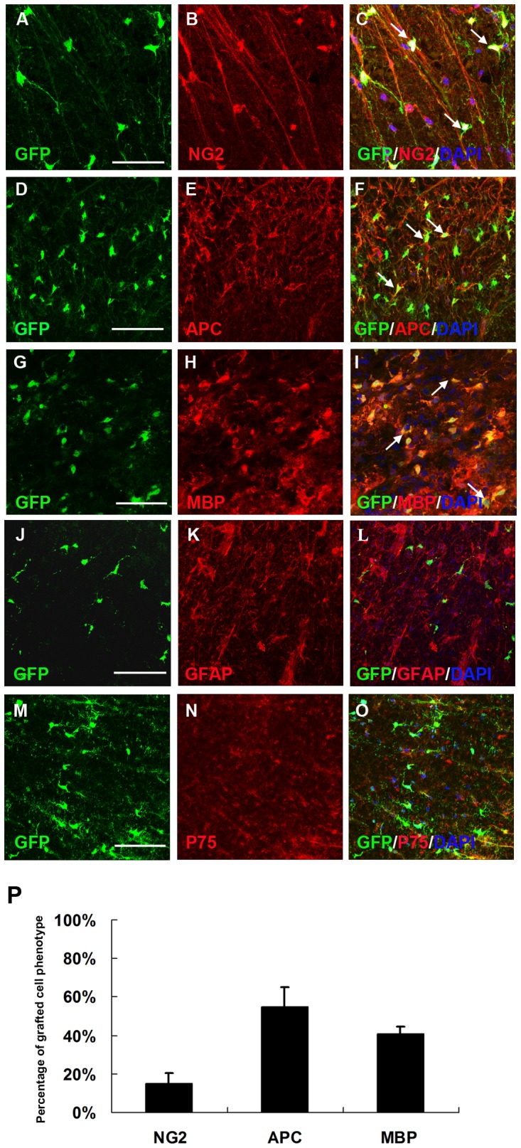
Figure 4. Olig2+-GFP+-OPCs primarily differentiate along the oligodendrocyte lineage.

At eight weeks after transplantation, a certain number of grafted GFP+ cells still expressed NG2 (A–C, arrows) and most of the grafted GFP+ cells became APC-positive mature oligodendrocytes (D–F, arrows). Double staining for GFP and MBP in cross-sections further confirmed that GFP-immunoreactive rings were composed of MBP+ myelin (G–I, arrows). The grafted GFP+ cells did not co-expressed GFAP (J–L) or P75 (M–O). (P) Quantification of GFP+ cell populations in the spinal cord. Data are expressed as the mean number of cells/spinal cord±SEM (n = 6). GFP+ cells expressed NG2 (15.1±5.53%), APC (54.6±10.5%) and MBP (40.5±3.8%). Bar, 50 µm (A–C); bar, 35 µm (D–O).
