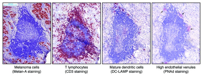
Figure 1. Microscopic images of an ectopic lymphoid structure in a metastatic melanoma lesion. Sequential cryosections from the tumor were immunostained against the indicated antigens and counterstained with hematoxylin. Positively stained antigens appear in red. The central structure with dense blue nuclei in the center of the images is a B-cell follicle, and the central pale structure is a germinal center. A T-cell area associating mature dendritic cells, T cells aggregates and high endothelial venules can be observed on top of the follicle.
