
Figure 1.
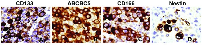
Immunohistochemical expression of CD133, ABCB5, CD166 and Nestin (A, B, C and D respectively) in FFPE tissues from primary melanoma. FFPE tissues were deparaffinized, and immunostained using a Bond-Max automated staining system (Leica). The reactions were developed using a biotin-free bond polymer refine detection kit (Leica) and visualized with 3′3-diaminobenzidine (DAB) substrate. Among positive immunostained tissues of primary melanomas, staining tended to be focal with a spectrum ranging from moderate-to-strong (original magnification x40).