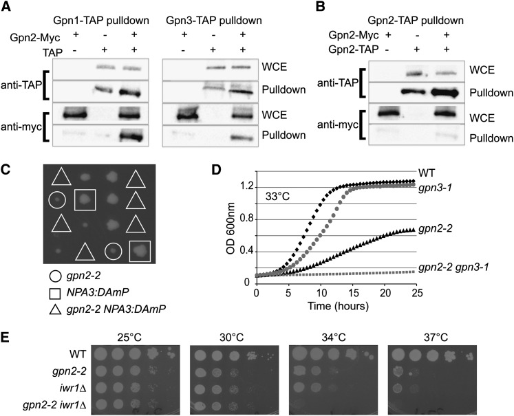
Figure 2.
Functional relationships between GPN proteins and Iwr1. Coprecipitation tests of Gpn2–13-Myc by TAP fusions of Npa3 or Gpn3 (A) or Gpn2–TAP (B) as measured by Western blotting with anti-TAP or anti-Myc antibodies. WCE, whole cell extract. (C, D, and E) Genetic interactions between GPN2 mutants and NPA3/GPN1, GPN3, and IWR1 mutants. (C) Tetrad dissection of gpn2-2 NPA3::DAMP double mutants. Triangles indicate where double mutant colonies should grow. (D) Growth curve assay of gpn2-2, gpn3-1, and double mutants at the indicated temperature. (E) Spot dilution assays of WT, gpn2-2, iwr1Δ, and double mutant cells. Cells were grown for 2 days at the indicated temperature.

