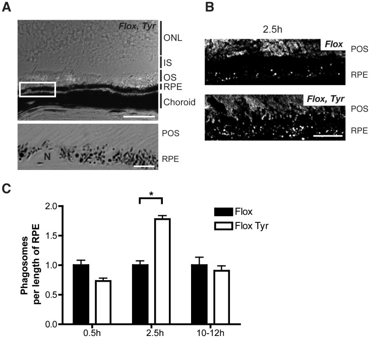
Figure 2. Increased number of phagosomes in ChmFlox, Tyr-Cre+ mice.
Frozen sections of eyes from 6-month old ChmFlox, Tyr-Cre+ mice and littermates ChmFlox mice harvested at the indicated times after light onset were immunostained with antibody (RetP1) for rhodopsin and analysed by confocal microscopy. Phase images were used to identify the RPE within the tissue. (A) Overview of the choroid, RPE, and part of the retina in ChmFlox, Tyr-Cre+ sample. Scale bar: 50 µm. The boxed region is magnified in the panel beneath and shows an area of the RPE with overlaying POS, scale bar: 5 µm. (B) Projections of 9 confocal sections. Scale bar: 10 µm. (C) Phagosomes were counted and results were normalised to the control and are presented as mean+/−SEM of 3–5 observations. *P<0.05. (ONL) Outer nuclear layer, (IS) Inner segment, (OS) Outer segment.

