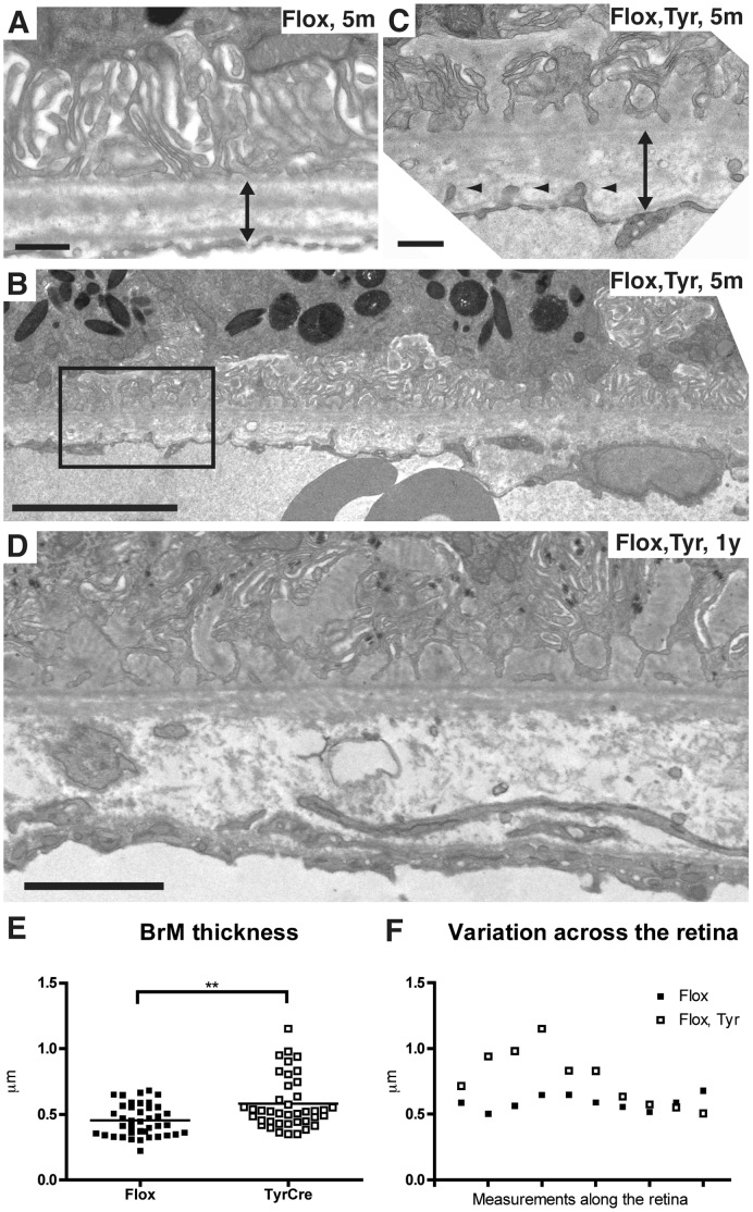
Figure 6. Thickening and abnormalities of Bruch’s Membrane in ChmFlox, Tyr-Cre+ mice.
Electron micrographs of 5-month old ChmFlox (A), littermate ChmFlox, Tyr-Cre+ (B–C) and 1-year old ChmFlox, Tyr-Cre+ mice (D). An enlargement of the box in B is shown in C. In ChmFlox, Tyr-Cre+ mice BrM becomes thicker with time. Double arrows show BrM thickness, small arrowheads indicate endothelial cell protrusions into BrM. Scale bars: 0.5 µm (A and C), 2 µm (B), 1 µm (D). (E) BrM thickness was measured in four 7-month old ChmFlox, Tyr-Cre+ mice (black square) and their littermate controls (grey dots). In each mouse 10 areas of retina were analysed. The two means are significantly different. ***P = 0.009. (F) Example of the variation of the measurements of BrM thickness along the retina for one ChmFlox, Tyr-Cre+ mouse (black square) and its littermate control (grey dots).

