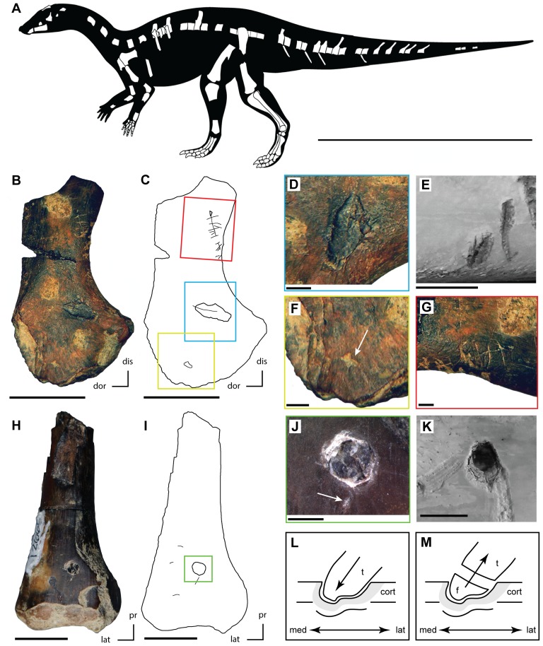
Figure 2. Feeding traces on juvenile ‘hypsilophodontid’ bones (Kaiparowits Formation) compared to those derived via actualistic experiments.
A. Skeletal reconstruction of the undescribed ‘hypsilophodontid’ from the Kaiparowits Formation with known material shown in white (modified from [65]). B. Partial left scapula (UMNH VP 21104) with feeding traces collected from UMNH locality 303. C. Outline drawing of left scapula (UMNH VP 21104) with feeding traces highlighted and colored boxes showing the locations of figure parts D, F, and G (colors match the respective figure parts). D. Bisected pit on the left scapula (UMNH VP 21104). E. Bisected pit on a modern cow femur produced by Alligator mississippiensis during actualistic experiments [20]. F. Small pit (highlighted by white arrow) on the proximal portion of the left scapula (UMNH VP 21104). G. Series of small scores present along the ventral margin of the neck of the left scapula (UMNH VP 21104). H. Distal portion of a right femur (UMNH VP 21107) with feeding traces collected from UMNH locality 303. I. Outline of right femur (UMNH VP 21107) with feeding traces highlighted and colored box showing the location of figure part J. J. Puncture containing an embedded tooth present on the right femur (UMNH VP 21107) and a small pit (highlighted by white arrow) just ventral to the puncture. K. Puncture present on a modern cow femur produced by A. mississippiensis during actualistic experiments [20]. L. Reconstruction of the hypothesized impact of the crocodyliform tooth with the right femur, creating the puncture observed in UMNH VP 21107. M. Reconstruction of the hypothesized fracturing of the damaged crocodyliform tooth crown, resulting in the embedded tooth observed in UMNH VP 21107. Scale bar equals one meter in A, 10 mm in B, E, H, and K, 2 mm in D, F, G, and J. Abbreviations: cort, cortical bone; dis, distal; dor, dorsal; f, tooth fragment; lat, lateral; med, medial; pr, proximal; t, tooth crown.

