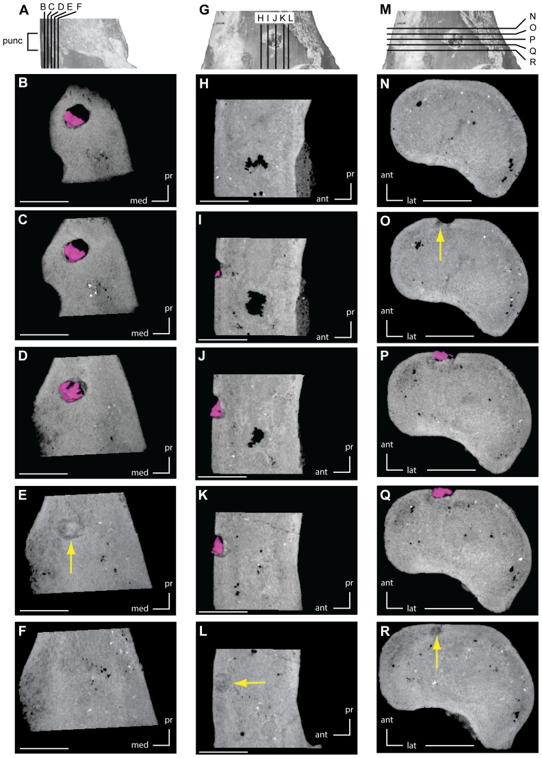
Figure 3. CT images of embedded tooth fragment and associated puncture on right femur (UMNH VP 21107).
A. Right femur in medial view showing orientation of images B–F. B–F. CT images taken along the coronal plane, arranged in order from anterior to posterior. G. Right femur in anterior view showing orientation of images H–L. H–L. CT images taken along the sagittal plane, arranged in order from lateral to medial. M. Right femur in anterior view showing orientation of images N–R. N–R. CT images taken along the transverse plane, arranged in order from proximal to distal. Pink areas outline the embedded tooth fragment, while yellow areas indicate regions of compression damage to the surrounding bone. Scale bars equal 5 mm. Abbreviations: ant, anterior; lat, lateral; med, medial; pr, proximal; punc, puncture.

