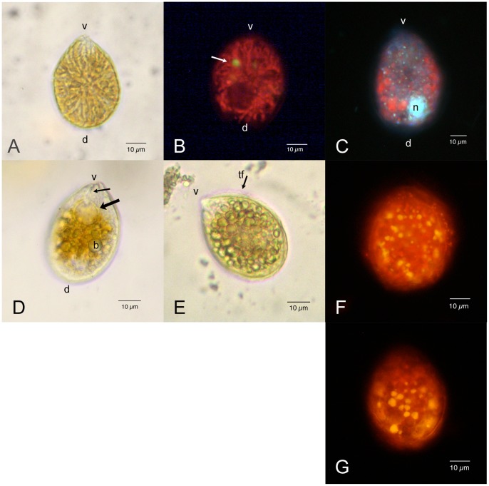
Figure 1. Ostreopsis cf. Ovata: Light and epifluorescence microscopy.
Scale bars represent 10 µm. (A) Living cell: bright field microscopy. The cell body is anterio-posteriorly compressed and shows a typical oval tear-shaped morphology pointed to the ventral side (v). The dorsal side (d) is rounded. Numerous elongated yellow brownish chloroplasts radiate from the centre of the cell. (B) Living cell viewed by epifluorescence microscopy under blue excitation (exciter filter BP 450–490, barrier filter LP 515): chloroplasts show an intense red autofluorescence; a small rounded body with yellow autofluorescence (arrow) is also visible. The dark non fluorescent area in the dorsal part of the cell is occupied by the nucleus. (C) Formaldehyde fixed cell stained with DAPI viewed by epifluorescence microscopy under UV excitation (exciter filter BP 340–380, barrier filter LP 425). The nucleus (n) occupies the dorsal part of the cell. Numerous small fluorescent dots (probably chloroplast DNA) are visible in correspondence with chloroplasts, which show a weak red autofluorescence. (D) Living cell: bright field microscopy. It can be observed the pusule (arrow), connected by a narrow canal (small arrow) to the cell ventral end (v). Some rounded translucent bodies (b) are evident in the cytoplasm. (E) Living cell: bright field microscopy. Most cytoplasm appears to be occupied by many rounded translucent bodies, which obscure other cell structures. It is visible the transverse flagellum (tf) running around the cell in the girdle. (F) Exponentially growing cell stained with Nile Red viewed by epifluorescence microscopy under blue excitation. Numerous yellow fluorescent lipid droplets are present in the peripheral cytoplasm. (G) Stationary phase cell stained with Nile Red. Yellow fluorescent lipid droplets appear to be larger.

