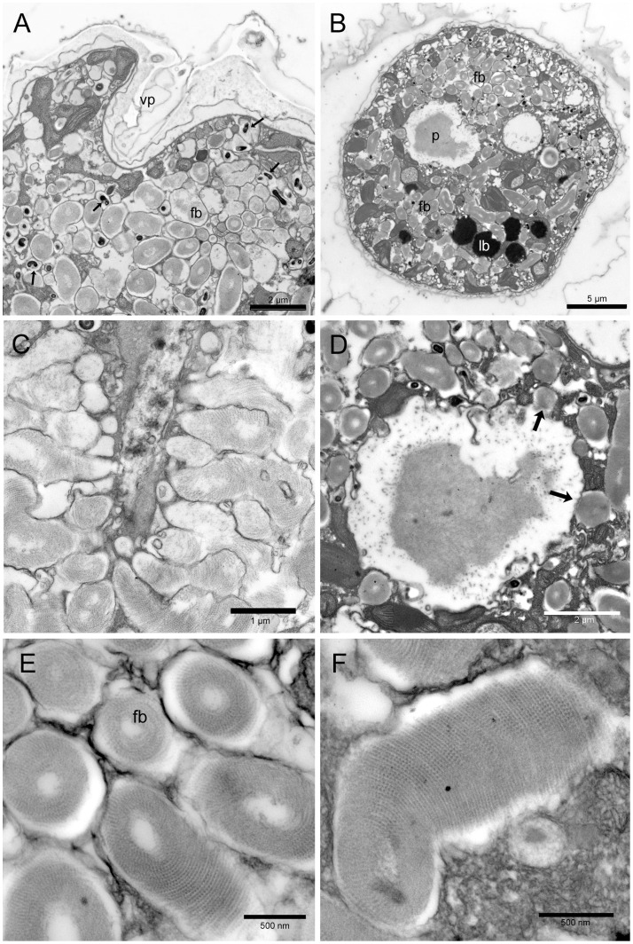
Figure 4. Ostreopsis cf. ovata fibrous bodies and pusule: Transmission electron microscopy.
(A) Oblique section of the ventral end of a cell showing the ventral pore (vp) and numerous fibrous bodies (fb) which occupy most cytoplasm. Smaller more electron dense fibrous bodies are also visible (arrows). Fixation 1. Scale bar 2 µm. (B) Transverse section of the cell showing the pusule chamber (p), containing electron dense material, surrounded by numerous fibrous bodies (fb). Lipid bodies (lb) are also visible. Fixation 1. Scale bar 5 µm. (C) Section of an elongated structure (possibly the pusule canal) with numerous fibrous bodies perpendicular to it: some of them seem to discharge their content into the canal. Fixation 2. Scale bar 1 µm. (D) Transverse section of the pusule chamber containing electron dense fibrous and amorphous material. Some fibrous bodies seem to discharge their content into the chamber (arrows). Fixation 1. Scale bar 2 µm. (E) Fibrous bodies (fb) in transverse and oblique section: they show an organized structure made of spirally arranged filaments packed around an electron transparent core. Fixation 1. Scale bar 500 nm. (F) Fibrous body in longitudinal section. Fixation 1. Scale bar 500 nm.

